| 1st | U | C | A | G | 3rd |
|---|
How to read the chart
Read a codon 5′ to 3′, one base at a time. The first base picks the row block, the second base picks the column, and the third base picks the line inside that cell. So for AUG you go to the A block, the U column, then the G line, and land on methionine. Reading a codon backwards gives a different amino acid entirely, so direction matters.
Notice how much of the redundancy sits in the third base. In many cells all four third-base options give the same amino acid, which is why that spot is called the wobble position and why a mutation there is so often silent. Leucine, serine, and arginine each get six codons, while methionine and tryptophan get only one apiece, which makes those two the most vulnerable to a single-base change.
Related tools: DNA → protein translation (translate a whole sequence) · Amino acid chart (all 20) · DNA melting temp & GC · Gene expression regulation · Biochem & MCAT equation sheet · all biochem tools.
The numbers worth memorizing
| Fact | Value | Why it matters |
|---|---|---|
| Total codons | 64 (43) | Three bases, four options each |
| Coding for amino acids | 61 | 64 minus the three stops |
| Start codon | AUG (Met) | Sets the reading frame; also codes Met mid-sequence |
| Stop codons | UAA, UAG, UGA | Read by release factors, not tRNA |
| Most codons | Leu, Ser, Arg (6 each) | Heavily buffered against mutation |
| Fewest codons | Met, Trp (1 each) | Any change alters the protein |
The code is degenerate (most amino acids have several codons) but not ambiguous (each codon specifies exactly one amino acid). It is also nearly universal, which is what makes it possible to express a human gene in bacteria.
Printable genetic code chart
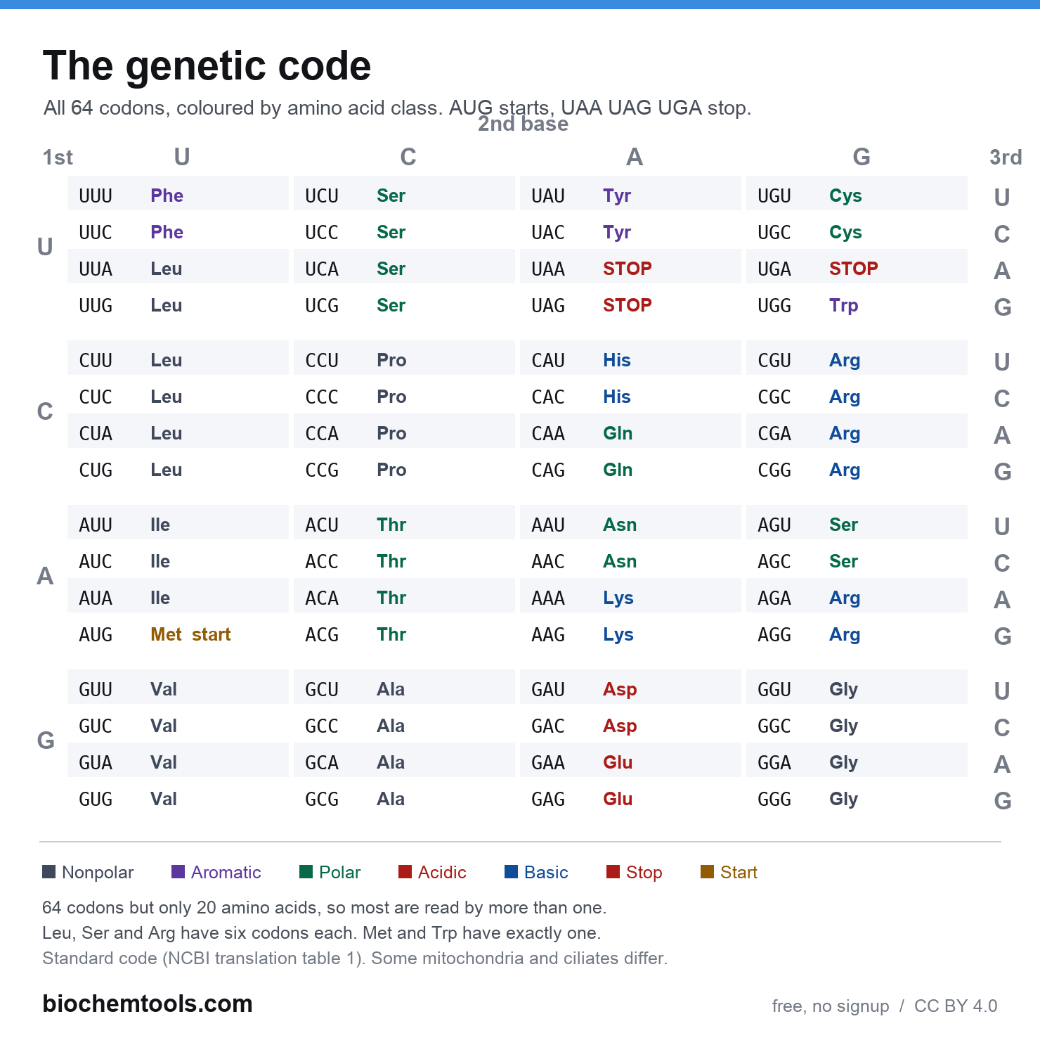
The same 64 codons as a single page you can print or keep on your phone, laid out in the classic first base / second base / third base grid and colour coded by amino acid class. Start and stop codons are marked.
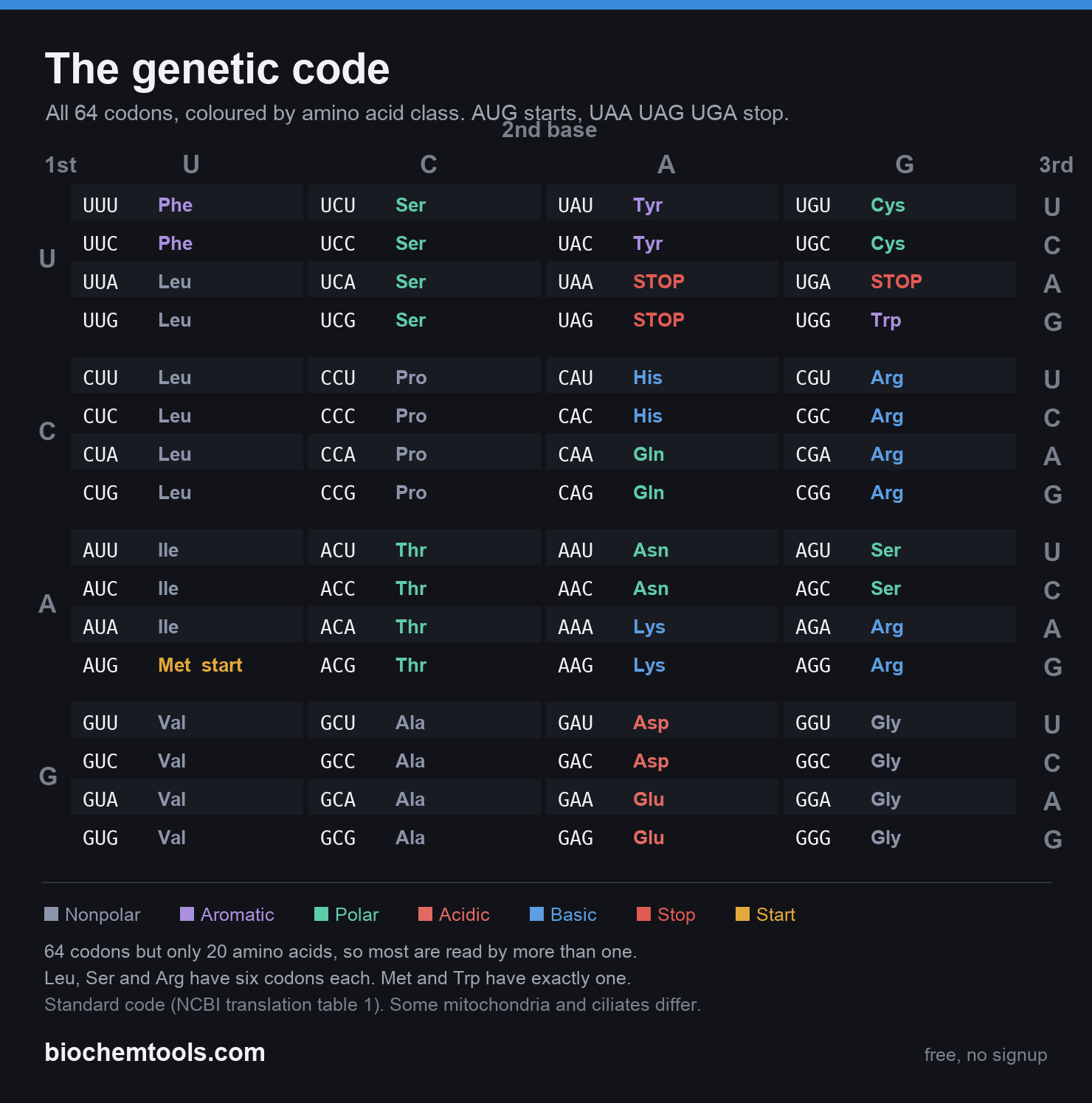
Download the chart, or the print-friendly white version. Free to use in a study guide or a class handout, no attribution needed. See all the printable charts. Codon assignments follow NCBI translation table 1 and were checked against an independently generated copy of the standard code.
FAQ
How do you read a codon chart?
5′ to 3′: first base = row block, second = column, third = line in the cell. AUG lands on methionine.
What are the start and stop codons?
Start: AUG (also Met). Stops: UAA, UAG, UGA, which code for no amino acid and are read by release factors.
What does degenerate mean?
Most amino acids have more than one codon (Leu, Ser, Arg have six; Met and Trp have one). Each codon still means exactly one thing.
What is the wobble position?
The third base, where tRNA pairing is looser, so one tRNA can read several codons and third-base mutations are often silent.
DNA vs RNA chart?
Same information, different letter: RNA's U is DNA's T, so AUG appears as ATG on the DNA coding strand. Use the toggle above.
Sources and how to cite this page
This chart is the standard genetic code, the code used by the nuclear genome of humans and almost all other organisms. It corresponds to NCBI translation table 1. AUG codes for methionine and serves as the start codon, and UAA, UAG, and UGA are the three stop codons. The amino acid class colours follow the usual side-chain grouping: nonpolar, aromatic, polar uncharged, acidic, and basic.
Where this code does not apply. A number of genomes use variant codes. Vertebrate mitochondria, for instance, read UGA as tryptophan rather than stop and AGA and AGG as stop rather than arginine. Several ciliates reassign UAA and UAG to glutamine. If you are working on mitochondrial or protist sequence, check which translation table applies before using this chart.