| Code ▲ | 3-letter | Name | Class | pKa COOH | pKa NH3+ | pKa R | pI | MW | Hydro. | Essential |
|---|
How to read this chart
Every amino acid shares the same backbone: an α-carboxyl group (pKa around 2) and an α-amino group (pKa around 9). What makes each one different is the side chain, and that side chain sets the class, the hydropathy, and whether the residue can carry a charge. Only seven of the 20 have an ionizable side chain (Asp, Glu, His, Cys, Tyr, Lys, Arg), and those seven are the only ones that affect a protein's net charge or its isoelectric point. The pI is the pH at which the molecule has no net charge, found by averaging the two pKa values that flank the neutral form: the two lowest for an acidic amino acid, the two highest for a basic one, and simply the backbone pair for the rest.
Related tools: Peptide charge & pI calculator · Amino acid titration curve · Amino acid quiz · Protein MW & extinction coefficient · DNA → protein translation · Codon chart (genetic code) · Biochem & MCAT equation sheet · Buffer pKa table · all biochem tools.
The seven ionizable side chains
These are the ones worth memorizing, because they are the only side chains that change charge in the normal pH range. Everything about protein charge, pI, buffering, and enzyme catalysis comes back to this short list.
A mnemonic that also gives you the order
“Don't Express Hate, Create Your Kindness Right” spells D E H C Y K R, which is all seven ionizable side chains listed in ascending pKa order, from aspartate at 3.65 up to arginine at 12.48. So the mnemonic gives you the list and the ranking at the same time, which is the part that usually matters in a charge question.
Thanks to u/The_528_Express on r/MCAT for this one.
| Amino acid | Side-chain pKa | Charge at pH 7 | Why it matters |
|---|---|---|---|
| Aspartate (D) | 3.65 | Negative | Deprotonated well below pH 7 |
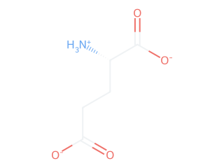
| Glutamate (E) | 4.25 | Negative | Deprotonated well below pH 7 |
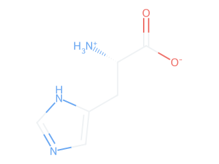
| Histidine (H) | 6.00 | Slightly positive | Only side chain that buffers near physiological pH; common in enzyme active sites |
| Cysteine (C) | 8.18 | Neutral (protonated) | Forms disulfide bonds when oxidized |
| Tyrosine (Y) | 10.07 | Neutral (protonated) | Absorbs at 280 nm, used to quantify protein |
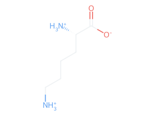
| Lysine (K) | 10.53 | Positive | Protonated far above pH 7 |
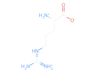
| Arginine (R) | 12.48 | Positive | Most basic side chain; essentially always positive |
Rule of thumb: an acidic group is charged (negative) when the pH is above its pKa; a basic group is charged (positive) when the pH is below its pKa.
All 20 structures
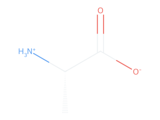
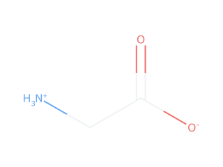
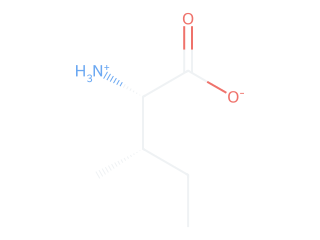
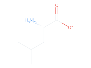
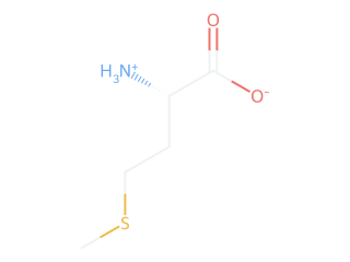
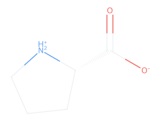
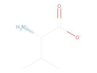
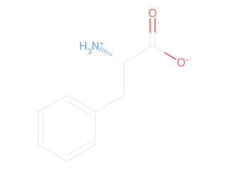
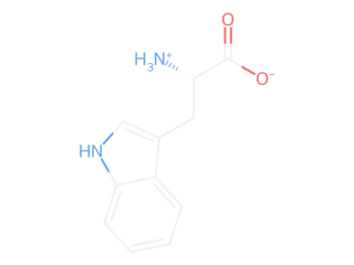
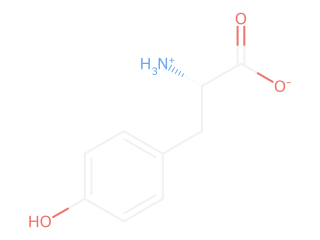
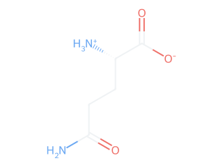
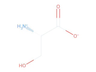
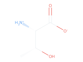
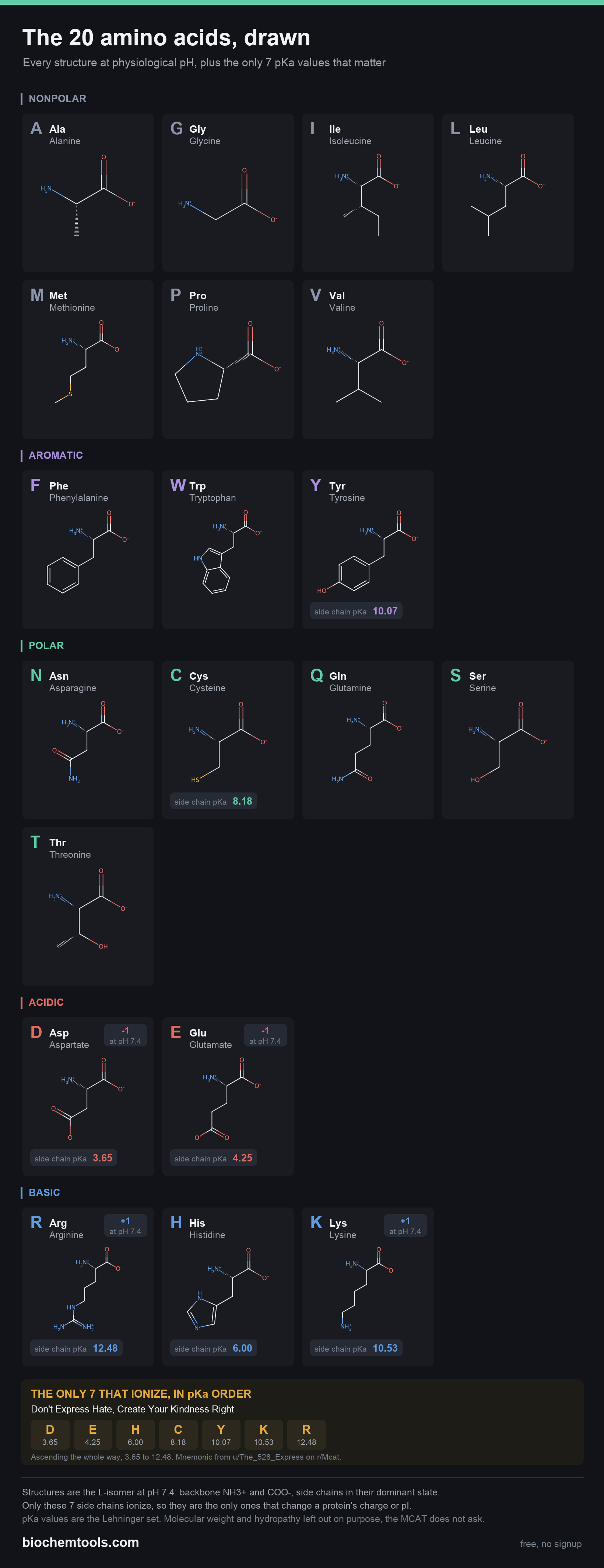
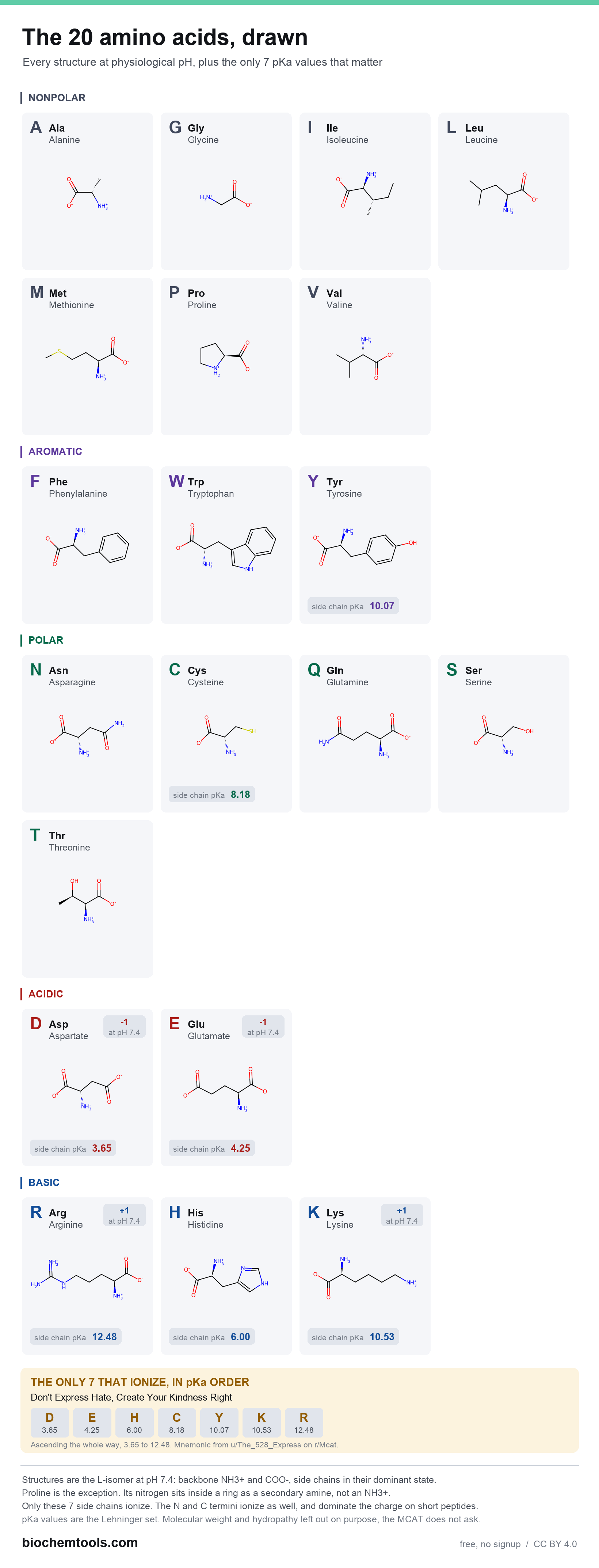
Every amino acid drawn as it actually exists at physiological pH: the backbone carries a positive α-amino group and a negative α-carboxyl group, and each side chain is shown in the form it takes at pH 7.4. All 20 are drawn on the same backbone orientation, so you can compare side chains by looking at the same spot on each one. Only the seven ionizable side chains list a pKa, because they are the only ones that can change.
Nonpolar
Aromatic
Polar
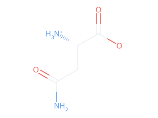
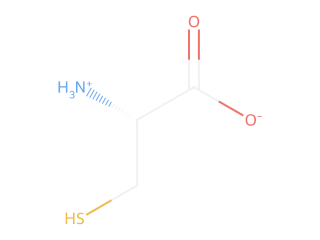
Acidic
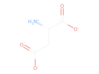
Basic
Want it on paper? Download the one-page structure chart, or the print-friendly white version (PNG, prints on a single sheet). Molecular weight and hydropathy are deliberately left off that version, since they are not what structure questions test.
FAQ
How are the 20 amino acids classified?
By side chain: nonpolar (G, A, V, L, I, P, M), aromatic (F, W, Y), polar uncharged (S, T, C, N, Q), acidic (D, E), and basic (K, R, H).
Which have ionizable side chains?
Seven: Asp 3.65, Glu 4.25, His 6.0, Cys 8.18, Tyr 10.07, Lys 10.53, Arg 12.48. Only these change a protein's net charge.
What is pI?
The pH of zero net charge. Average the two pKa values flanking the neutral form: the two lowest for acidic, two highest for basic, backbone pair otherwise.
Which are essential?
Nine: His, Ile, Leu, Lys, Met, Phe, Thr, Trp, Val. Six more (Arg, Cys, Gln, Gly, Pro, Tyr) are conditionally essential.
What is hydropathy?
The Kyte-Doolittle index: positive is hydrophobic (Ile +4.5 highest), negative is hydrophilic (Arg −4.5 lowest). Predicts buried residues and membrane helices.
Sources and how to cite this page
The pKa and pI values in this chart are the standard set tabulated in Lehninger Principles of Biochemistry (Nelson & Cox), which is the set used in most biochemistry courses and on the MCAT. Molecular weights are residue masses plus water, computed from atomic masses and cross-checked against the free amino acid formula weights. The hydropathy column is the Kyte-Doolittle scale (Kyte, J. & Doolittle, R.F., Journal of Molecular Biology 157(1):105–132, 1982).
Why published pKa values differ slightly. pKa is measured, not derived, so a tabulated value depends on temperature and ionic strength. This table, like most, is for dilute aqueous solution near 25 °C. Differences of roughly 0.1 to 0.3 pKa units between textbooks are normal and do not mean one of them is wrong. Inside a folded protein the gap can be much larger, because nearby charges and the local dielectric shift a side chain well away from its free-amino-acid value. If you are citing a number for coursework, cite the source you were taught from and keep it consistent.